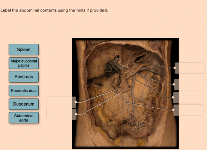
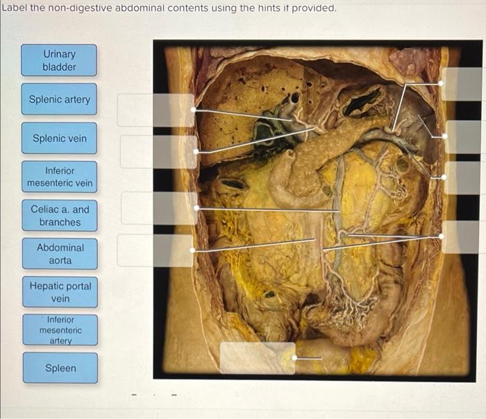
Label the abdominal contents using the hints if provided. – Labeling the abdominal contents using the hints provided is a crucial aspect of understanding abdominal anatomy. This comprehensive guide will provide a detailed explanation of the importance of accurate labeling, a table organizing the abdominal contents and their corresponding labels, and hints for effective labeling.
By understanding the abdominal contents and their relationships, healthcare professionals can gain a deeper comprehension of abdominal anatomy and its clinical implications.
The abdominal cavity houses a multitude of vital organs and structures, each with specific functions and anatomical relationships. This guide will delve into the major organs and structures within the abdominal cavity, exploring their functions and interconnections. Additionally, the four quadrants and nine regions of the abdomen will be defined, highlighting their clinical significance.
Labeling Abdominal Contents: Label The Abdominal Contents Using The Hints If Provided.

Accurately labeling abdominal contents is crucial for effective medical diagnosis, treatment, and surgical procedures. A comprehensive understanding of the anatomical structures within the abdomen aids in precise localization and manipulation of organs during medical interventions.
The following table provides a systematic approach to labeling the abdominal contents, along with hints for accurate identification:
| Structure | Label | Hint |
|---|---|---|
| Liver | L | Largest organ in the abdomen |
| Stomach | S | J-shaped organ located in the upper left quadrant |
| Small intestine | SI | Long, coiled tube responsible for nutrient absorption |
| Large intestine | LI | Colon and rectum; involved in waste elimination |
| Pancreas | P | Located behind the stomach, produces digestive enzymes |
| Gallbladder | GB | Small, pear-shaped organ attached to the liver |
| Spleen | SP | Located in the upper left quadrant, filters blood |
| Kidneys | K | Bean-shaped organs responsible for urine production |
Identifying Organs and Structures within the Abdominal Cavity

The abdominal cavity houses numerous vital organs and structures, each performing distinct functions:
- Liver:Largest abdominal organ, involved in metabolism, detoxification, and bile production
- Stomach:J-shaped organ where food is broken down by digestive enzymes
- Small intestine:Coiled tube responsible for nutrient absorption and digestion
- Large intestine:Colon and rectum, involved in waste elimination and water absorption
- Pancreas:Produces digestive enzymes and hormones, including insulin
- Gallbladder:Stores and releases bile for fat digestion
- Spleen:Filters blood and stores red blood cells
- Kidneys:Filter blood and produce urine
- Ureters:Tubes that carry urine from the kidneys to the bladder
- Bladder:Stores urine before elimination
These organs are interconnected and work in harmony to maintain homeostasis and perform essential bodily functions.
Regions of the Abdomen

The abdomen is divided into four quadrants and nine regions to facilitate precise anatomical localization:
- Quadrants:
- Right upper quadrant (RUQ)
- Right lower quadrant (RLQ)
- Left upper quadrant (LUQ)
- Left lower quadrant (LLQ)
- Regions:
- Hypochondriac (right and left)
- Epigastric
- Lumbar (right and left)
- Inguinal (right and left)
- Hypogastric
Understanding these regions is crucial for accurate diagnosis and localization of pain or pathology within the abdomen.
Peritoneal Cavity and Its Role in Abdominal Anatomy
The peritoneal cavity is a fluid-filled space that lines the abdominal wall and covers the abdominal organs:
- Structure:Consists of two layers, the parietal peritoneum lining the abdominal wall and the visceral peritoneum covering the organs
- Function:Facilitates organ movement, provides lubrication, and contains immune cells
- Relationship with Organs:Organs are suspended within the peritoneal cavity by mesenteries, which are double-layered folds of peritoneum
Disorders of the peritoneal cavity, such as peritonitis, can lead to significant inflammation and complications.
Blood Supply and Innervation of Abdominal Contents

The abdominal contents receive blood supply from major arteries and veins:
- Arteries:
- Celiac trunk
- Superior mesenteric artery
- Inferior mesenteric artery
- Veins:
- Hepatic portal vein
- Superior mesenteric vein
- Inferior mesenteric vein
The abdominal organs are innervated by both sympathetic and parasympathetic nerves:
- Sympathetic:Originate from the thoracic and lumbar spinal cord, control organ function during stress
- Parasympathetic:Originate from the vagus nerve, control organ function during digestion and rest
Understanding the blood supply and innervation of the abdominal contents is essential for surgical interventions and management of abdominal disorders.
FAQ Overview
What are the key considerations for accurate labeling of abdominal contents?
Accuracy, consistency, and adherence to established anatomical terminology are essential for effective labeling.
How can hints enhance the labeling process?
Hints provide additional information or clues that can assist in identifying and labeling the abdominal contents.
What is the clinical significance of understanding the abdominal regions?
Understanding the abdominal regions aids in localizing pain, guiding diagnostic procedures, and planning surgical interventions.